Here are my eyes: looking in at some of the many eye diagnoses I have – and looking out at some of the gorgeous splendid amazing things I’ve seen.
Before my eyes changed, that is.
Im visually impaired – I can’t see bus numbers or street signs – or even my friends in the street. Much of what is online is very difficult for me to see.
But look at what I can create!
‘Impaired’ is maybe not quite the right word
Even if I’m ridiculously nose-to-screen when I’m making my creations!
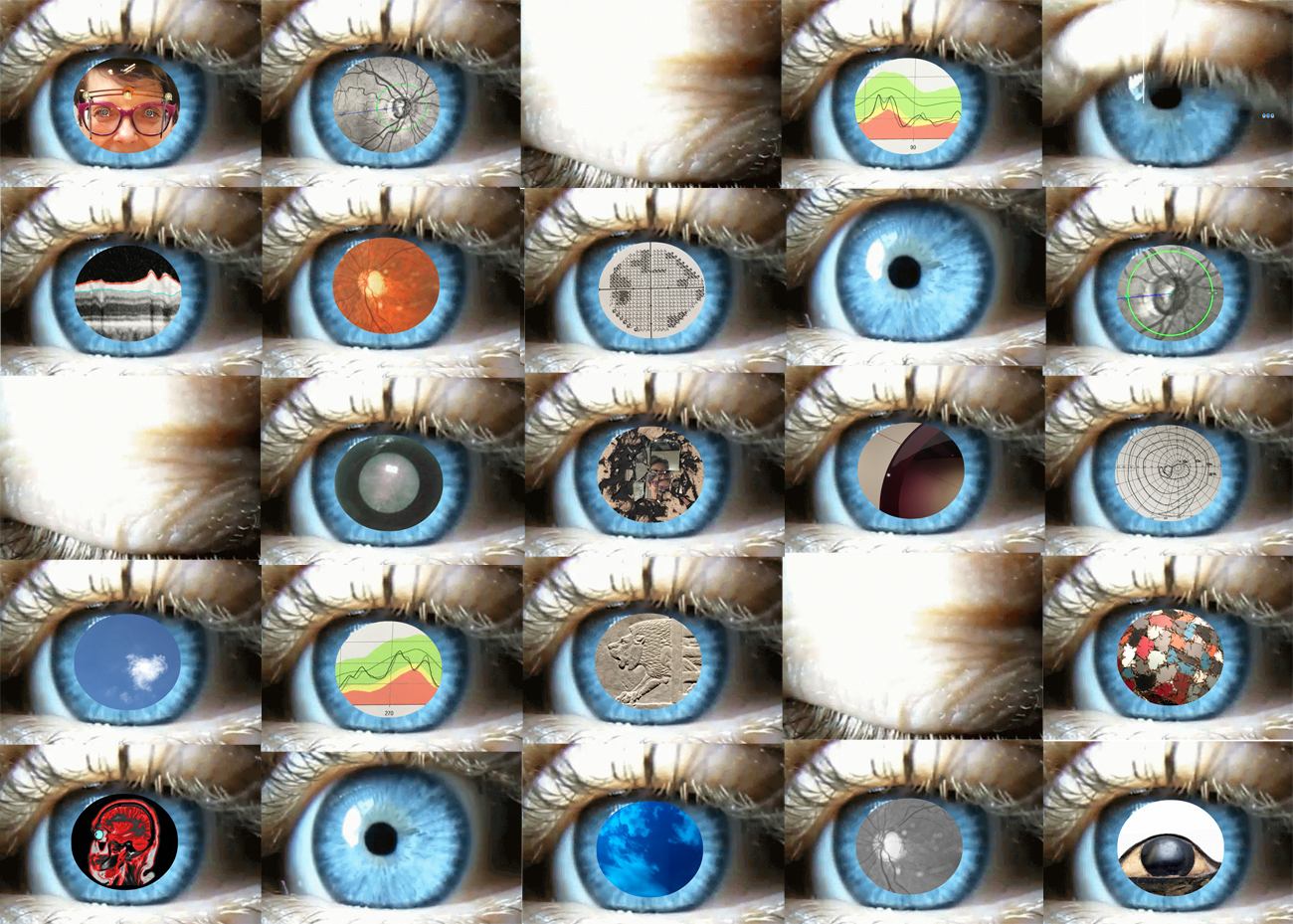
Here’s a description of what you might – or might not – be able to see.
There’s a five by five grid of eyes – mainly open, but with a few shut like in a blink.
The open eyes are blue, and with long upturned eyelashes.
Most of the open eyes have a dilated (enlarged) pupil.
Showing either an image of my eye looking in – to show some of the many different tests the many different eye-doctors have done on my eyes
Or an image looking out – to some of the many glorious things my eyes have seen.
In detail, left to right, top to bottom:
Top row:
1.1. Being measured for spectacles at an opticians
1.2. A black snd white photo of one of my retina – you can see the dark optic disc, with the arteries and veins swirling over it
1.3. A blinked eye-lid
1.4. An abnormal OCT (optical coherence tomogram). Green is normal, and red isn’t
1.5. Open eye, with undilated pupil. My eyes are no longer blue – they’re more hazel, or green (very happy) / grey (less happy). But I couldnt get a motion photo of my own eye – so i”‘ve borrowed someone else’s
Second row:
2.1. Looking around my eyes in black and white
2.2. Colour retinal photography
2.3. Abnormal visual field – the dark bits shouldn’t be there.
2.4. Open eye, with undilated pupil.
2.5. Black and white retinal photography. I think the green circle is about assessing the depth of the retina? Maybe?!
Third row:
3.1. A blinked eye-lid
3.2. Showing the start of lens opacification. The beginnings of cataracts is why I cant handle any dazzle. And wear antiglare shields.
3.3. Reflections in mirrored exhibit in the Hayward Gallery
3.4. Part of one of the machines use by the eye doctors. All these machines measuring things – but havent ever come up with any useful suggestions!
3.5. An old fashioned hand-drawn visual field
Fourth row:
4.1. A cloud seen near a piece on a terrace outside the Hayward gallery
4.2. A less abnormal OCT (optical coherence tomogram). With more normal Green and ;less abnormal red.
4.3. An Assyrian lion – in a stone relief
4.4. A blinked eye-lid
4.5. Detail of a patchwork ball – show in the Hayward Gellery
Fifth row:
5.1. Sagittal view (cut longways down the middle) of an abnormal brain scan- with MS-related demyelination
5.2. Open eye, with undilated pupil.
5.3. Clouds reflected in a parabolic mirror on a terrace outside the Hayward gallery
5.4. More black and white retinal photography. Are those dots my PVD?
5.5. This image is of one of the anatomically accurate eyes from an ancient Egyptian sculpture.
I only realise as I write this, how many of the looking-out images here are from the Hayward gallery. I had lots of other looking-out to outside-nature images – maybe they felt too big and unconstrained for my made-up pupils?